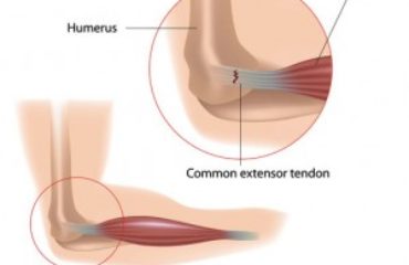
A fractured elbow essentially means the fracture of the olecranon process of the ulna. It’s the tip of the elbow that typically breaks in an elbow fracture as it is positioned directly under the skin of the elbow, without much intervening soft tissue. Therefore, it is susceptible to fracture as a result of direct trauma, typically when falling on an on an outstretched arm. A fractured elbow can be extremely painful and make elbow motion difficult or impossible. If the trauma is severe, it is possible to have concomitant shoulder injuries as well.
Treatment for an olecranon fracture depends upon its extent and severity. For simple fractures, wearing a splint may be enough. However, if it involves displaced pieces of bone, surgery is required most often required.
Physical examination
Diagnosing the fractured elbow and determining the extent of the injury depends on a focused physical examination of the elbow and the shoulder, assessing the amount of swelling, bruising, and to see if there is disfigurement, and taking a detailed history about the type and severity of pain. An open fracture is when the bone protrudes through the skin. These are serious injuries should be immediately treated with surgery.
Closed fractures are in which the bone fragments remain beneath the skin, examining for which requires performing a full range of motion so that the damage to bones, tendons, or ligaments can be assessed.

Diagnostic Imaging
Diagnostic workup starts with an X-ray, which can reveal the location of a fractured bone in the shoulder or elbow. It can also reveal if its a hairline, simple, or complex fracture and how displaced the bone fragments are, if at all. An x-ray is relatively cheap, easily available (usually at urgent cares and doctor’s offices) and delivers a small radiation dose.
A CT scan, on the other hand, involves more radiation and costs more, but delivers two- or three-dimensional computer images of the shoulder or elbow, allowing a much more detailed assessment of the extent of the injury.
It is usually considered when the X-ray reveals that the fracture is complex and/or with displaced bone fragments. Newer techniques have made CT scans safer as they deliver much smaller radiation doses.
MR imaging can be performed if the damage to the shoulder or elbow is severe and injury to adjacent ligaments, tendons, or nerves is suspected. An MRI scan uses magnetic fields to take detailed images of the soft tissue and can be especially helpful in the assessment of torn or strained ligaments or tendons, or if there is nerve impingement or damage. MRI scans don’t involve any radiation exposure but are more expensive to perform, which is why they are never the first-line diagnostic tests and are reserved for patients with severest suspected tissue injury. It does, however, have some contraindications, such as metallic implants or contrast allergies.


