Cartilage Injuries
The knee joint is composed of three bones: the kneecap (patella), thigh bone (femur), and shin bone (tibia).
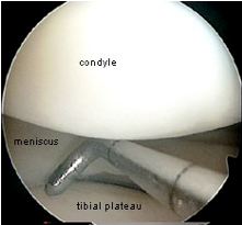
The end of each of these bones is covered by a smooth, shiny, white, Teflon-like layer of tissue called articular cartilage. This layer allows these bones to glide smoothly over each other during knee motion. The following two pictures show normal articular cartilage.
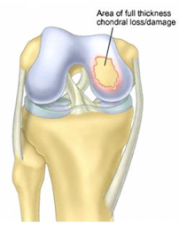
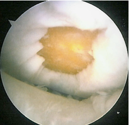
This articular cartilage can be damaged during a twisting or pivoting i njury, or by a direct impact to the knee. The following two pictures show a defect of the articular cartilage following an injury.
Common mechanisms of injury include falls, sports injuries, or motor vehicle accidents. Injuries to the articular cartilage, or chondral injuries, may also occur in association with ligamentous injuries, such as an ACL tear, or a meniscal injury (See ACL Injuries or Meniscal Tears).
Small pieces of the cartilage may occasionally break off and float around in the knee as loose bodies. Often, however, there is no clear history of a single injury. The patient’s condition may result from a series of minor injuries that have occurred over time.
Signs & Symptoms:
Although patients with a chondral injury may be asymptomatic, frequently knee pain is present. The pain is often worse with weight-bearing activities. Depending on the location of the injury within the knee, deep knee bending may exacerbate the pain. If the patella (knee cap) or trochlea of the femur is involved, symptoms may be worse with stairs and squatting.
In addition to pain, swelling in the knee may occur with chondral injuries.
Also, mechanical symptoms, such as locking, catching, or painful popping may occur. In patients with loose bodies, patients sometimes report the sensatio n that they feel something loose moving around in the knee.
Diagnosis:
Chondral injuries can be very difficult to diagnose after performing a thorough history and completing a comprehensive physical examination. Rarely, x-rays will be sufficient to make the diagnosis, but frequently the x-rays are normal.
An MRI scan will usually be sufficient to make the diagnosis; occasionally, however, a chondral lesion can be missed by the MRI, and is only detected at the time of surgery.
Treatment:
The optimal treatment for chondral injuries depends upon the size and location of the lesion, the age and activity level of the patient, the presence of a loose body, the symptoms experienced by the patient, and the presence or absence of arthritis in the remainder of the knee.
For older, low demand patients with minimal symptoms, non-operative treatment may be recommended. Non-operative treatment modalities include: weight loss, activity modification, anti-inflammatory medications, and steroid and/or viscosupplementation injections. These modalities may decrease the symptoms, but none of them will actually heal the defect/injury.
For younger, active patients or those who have failed non-operative treatment, surgery is often recommended. The type of surgical treatment depends upon numerous factors including patient age, the size and location of the lesion, and prior surgical treatments.
One commonly performed procedure, called a chondroplasty, occurs when the surgeon arthroscopically smoothes the shredded or frayed articular cartilage.
Another slightly more involved procedure, called microfracture, occurs when the surgeon performs arthroscopic surgery to create several small holes in the lesion to stimulate new cartilage growth.
To view a video animation depicting microfracture surgery, click the box below.
Open Surgery
For larger lesions, or for patients who have failed chondroplasty and/or microfracture, open surgical procedures may be considered, including:
– Osteochondral Autograft Resurfacing: Cartilage plugs are taken from other locations in the patient’s knee and are used to fill-in the void at the site of injury.
– Osteochondral Allograft Resurfacing: Cartilage plugs are taken from a donor cadaver knee and are used to fill-in the void at the site of injury.
– Autologous Chondrocyte Implantation: This is a two-staged procedure. In the 1st stage, arthroscopic surgery is performed to evaluate the chondral lesion and obtain a sample of the patient’s normal articular cartilage. This sample of cartilage is then sent to a laboratory where millions of cartilage cells are grown outside the body in tissue culture over a 3-6 week period. These cells can then be reimplanted into the injury site during the second procedure.
Dr. Farber Shows the Knee Microfracture Procedure

